Human Eye Dissection, Cow Eye Dissection : Notice the muscles surrounding the eye.. Carefully cut away the fat and the muscle. Join ross exton on a journey looking inside a horse eyeball, investigating the anatomy of this fascinating organ along the way.this vi. Diabetes and healthy eyes toolkit and website keywords: It also shows how various lenses can be used to correct for faulty vision. Differences between the two eye types will be mentioned as the dissection is completed.
Wear latex gloves if you have cuts in your hands. Now you want to remove the lens. Human anatomy diagrams show internal organs, cells, systems, conditions, symptoms and sickness information and/or tips for healthy living. Join ross exton on a journey looking inside a horse eyeball, investigating the anatomy of this fascinating organ along the way.this vi. The human eye and vision.
Instructions For Sheep Eye Dissection from s3.studylib.net The eye most likely has a thick covering of fat and muscle tissue. (just use youtube's search feature with these key words.) anatomy and function of the eye: These muscles move the eye up and down, side to side, and rotate the eye. Human eye dissection anatomy isolated on white background. Be aware that it is a simplified version of what actually happens. In the center of the retina is the optic nerve, a circular to oval white area measuring about 2 x 1.5 mm across. Professor, department chair, surgeon, neuroscientist and m. As you get closer to the actual eyeball, you may notice muscles that are attached directly to the sclera and along the optic nerve.
Parts of the eye outside the eyeball.
Place the preserved cow eye on a tray. At the exploratorium, we dissect cows' eyes to show people how an eye works. Human anatomy diagrams show internal organs, cells, systems, conditions, symptoms and sickness information and/or tips for healthy living. In the center of the retina is the optic nerve, a circular to oval white area measuring about 2 x 1.5 mm across. Begin the dissection by gathering the equipment and supplies listed here. National eye institute , national eye health education program subject: This is a simulation demonstrating the optics of the human eye. When surveyed about the five senses — sight, hearing, taste, smell and touch — people consistently report that their eyesight is the mode of perception they value (and fear losing) most. The human eye and vision. From the center of the optic nerve radiates. This diagram depicts eye dissection. Human eye dissection anatomy isolated on white background. Posted by raphael fernandez (2 minutes) human eye posted by smart learning for all (cartoon, 10 minutes) a journey through the human eye posted by bausch and lomb (2.5 minutes)
Parts of the eye outside the eyeball. Examine the back of the eye and find the optic nerve, a white, thick cord attached to the back of the eye. Notice the muscles surrounding the eye. This contribution explores both the form and function of numerous anatomic features of the human ocular system, which are vital to a comprehensive understanding of the pathophysiology of many oculocutaneous diseases. It's a clear lump about the size and shape of a squashed marble.
Eye Dissection Gcse A Level Biology Neet Practical Skills Youtube from i.ytimg.com Diabetes and healthy eyes toolkit and website keywords: Carefully cut away the fat and the muscle. In the center of the retina is the optic nerve, a circular to oval white area measuring about 2 x 1.5 mm across. Learn how to dissect a cow's eye in your classroom. How do your eyes work? Join ross exton on a journey looking inside a horse eyeball, investigating the anatomy of this fascinating organ along the way.this vi. Put on your personal protective equipment: 99 get it as soon as mon, jul 12
Parts of the eye outside the eyeball.
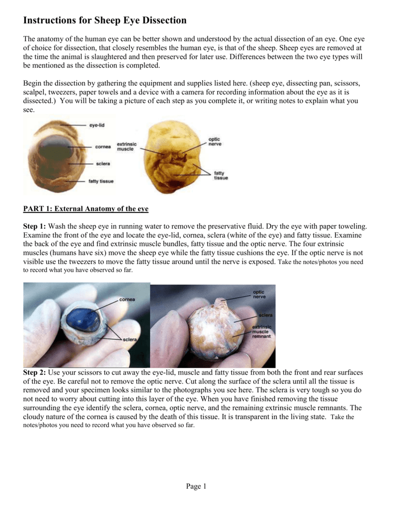
What is the purpose of the fatty tissue surrounding the eye? If you try this at home, wash your hands after the dissection. A closer look at the parts of the eye. The anatomy of the human eye can be better shown and understood by the actual dissection of an eye. Parts of the eye outside the eyeball. Mr exham is a biology teacher who likes making sense of biology. This diagram depicts eye dissection. Examine the external characteristics of the eye. Diabetes and healthy eyes toolkit and website keywords: One eye of choice for dissection, that closely resembles the human eye, is that of the sheep. These muscles move the eye up and down, side to side, and rotate the eye. 99 get it as soon as mon, jul 12 At the exploratorium, we dissect cows' eyes to show people how an eye works.
This web site shows photos and videos of a dissection. Place the cow eye on a dissecting tray. I run a blog designed to share resources for both students and teachers.this is a video of a. The main parts of the human eye are the cornea, iris, pupil, aqueous humor, lens, vitreous humor, retina, and optic nerve. Carefully cut away the fat and the muscle.
Human Eye Dissection Anatomy Brillen Galerie Koln from www.brillen-galerie-koeln.com The extraocular muscles are attached to the white part of the eye called the sclera. Learn how to dissect a cow's eye in your classroom. This web site shows photos and videos of a dissection. At the exploratorium, we dissect cows' eyes to show people how an eye works. Posted by raphael fernandez (2 minutes) human eye posted by smart learning for all (cartoon, 10 minutes) a journey through the human eye posted by bausch and lomb (2.5 minutes) It's a clear lump about the size and shape of a squashed marble. Human kidney magnification from a body as a medical diagram with a cross section of the inner organ with red and blue arteries and adrenal gland as a health care illustration of the anatomy of the urinary system. Wear latex gloves if you have cuts in your hands.
Mr exham is a biology teacher who likes making sense of biology.
The extraocular muscles are attached to the white part of the eye called the sclera. A closer look at the parts of the eye. Human eye dissection anatomy isolated on white background. It also shows how various lenses can be used to correct for faulty vision. Notice the muscles surrounding the eye. Differences between the two eye types will be mentioned as the dissection is completed. Diabetes and healthy eyes toolkit and website keywords: Wear latex gloves if you have cuts in your hands. National eye institute , national eye health education program subject: If you try this at home, wash your hands after the dissection. How do your eyes work? Human kidney magnification from a body as a medical diagram with a cross section of the inner organ with red and blue arteries and adrenal gland as a health care illustration of the anatomy of the urinary system. That's the vitreous humor, a mixture of protein and water.
Share
Post a Comment
for "Human Eye Dissection, Cow Eye Dissection : Notice the muscles surrounding the eye."


Post a Comment for "Human Eye Dissection, Cow Eye Dissection : Notice the muscles surrounding the eye."